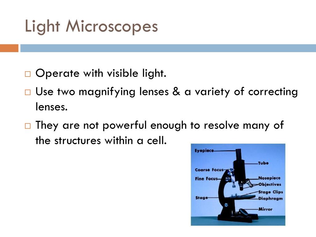

Samples for light microscopy are prepared in an ever-increasing number of techniques, and can range from sliced biological organisms and tissue.

Examples of light microscopes include brightfield microscopes, darkfield microscopes, phase-contrast microscopes, differential interference contrast microscopes, fluorescence microscopes, confocal scanning laser microscopes, and two-photon microscopes. They can be used as a primary visualization tool or in support of electron microscopy. Many types of microscopes fall under the category of light microscopes, which use light to visualize images. (Color online) Depolarized transmission light microscopy (DTLM) images of a liquid crystal cell at topographic line patterns. Among other things, proper care of a microscope includes the following: Light microscopes play an important role in many research laboratories, including electron microscopy facilities. In addition, microscopes are rather delicate instruments, and great care must be taken to avoid damaging parts and surfaces. Electrons have a shorter wavelength in comparison to light which. The major difference is that light microscopes use light rays to focus and produce an image while the TEM uses a beam of electrons to focus on the specimen, to produce an image.

#Transmission light microscope images skin#
In this figure An example of using two-photon microscopy to see blood vessels through the skin (few mm thickness) of a living mouse. The working principle of the Transmission Electron Microscope (TEM) is similar to the light microscope. Since lenses are carefully designed and manufactured to refract light with a high degree of precision, even a slightly dirty or scratched lens will refract light in unintended ways, degrading the image of the specimen. Light-sheet fluorescence microscopy can acquire images at speeds 100 to 1000 times faster than traditional confocal microscopes, which is very useful to study living cells. Without immersion oil, light scatters as it passes through the air above the slide, degrading the resolution of the image.Įven a very powerful microscope cannot deliver high-resolution images if it is not properly cleaned and maintained. (b) Because immersion oil and glass have very similar refractive indices, there is a minimal amount of refraction before the light reaches the lens. Its got loads of relevant information, very realistic simulators and. \( \newcommand\): (a) Oil immersion lenses like this one are used to improve resolution. MyScope is a fantastic online training tool for microscopy and microanalysis.


 0 kommentar(er)
